
- Haber Akışı
- KEŞFEDIN
- Sayfalar
- Etkinlikler
- Bloglar
-
 Hakkımızda
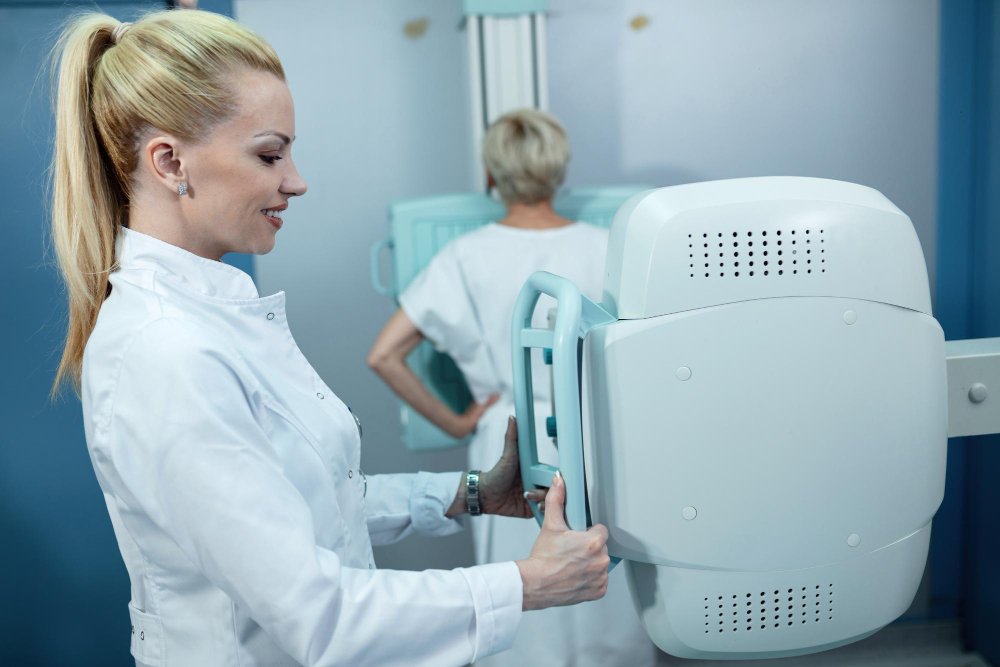
3D Breast CT and the Future of Breast Cancer Detection
Breast cancer screening in the United States is at an interesting inflection point. The tools we've been using for decades have saved lives — that's not in question. But the limitations of those tools are well-documented, and the research community has spent years developing approaches that address those limitations in meaningful ways.
3D breast CT is one of the most promising of those advances. It's not a headline-grabbing overnight breakthrough — it's the result of sustained engineering and clinical research effort over more than a decade, progressively refining a technology until it reaches the point where it can genuinely change what's possible in breast imaging. Understanding where that effort has led, and what it means for patients and providers navigating breast health decisions today, is worth the time.
Why Breast Imaging Keeps Evolving
The history of breast imaging is essentially a story of iterative problem-solving. Film-screen mammography was a major advance when it became standard of care. Digital mammography improved image quality and reduced dose. Tomosynthesis addressed the tissue overlap limitation that film and digital mammography shared. Each advance addressed real problems — and each revealed new ones or left prior limitations only partially resolved.
The persistence of false negatives — real cancers that imaging misses — and false positives — callbacks and biopsies for findings that turn out to be benign — drives continued investment in better tools. The consequences of these errors are different in nature but equally real. A missed cancer delays treatment during a window when treatment is most effective. An unnecessary biopsy creates anxiety, cost, and physical discomfort for a patient who didn't need the intervention.
Improving the sensitivity and specificity of breast imaging simultaneously — catching more real cancers while generating fewer false alarms — is the goal that motivates the development of technologies like 3D breast CT. And the early evidence suggests this technology moves meaningfully in both directions.
The Technical Foundations of Dedicated Breast CT
Understanding what makes dedicated 3D breast CT different from other imaging approaches requires a brief look at the underlying technology.
The fundamental challenge in designing a dedicated breast CT system is capturing a complete volumetric image of the breast at adequate resolution and acceptable radiation dose. The breast is not the easiest structure to image — it's mobile, its density varies substantially across patients, and it contains structures at multiple size scales that are all clinically relevant, from microcalcifications measured in fractions of a millimeter to masses measured in centimeters.
Current dedicated breast CT systems address this challenge with detector arrays that provide high spatial resolution across the full breast volume, X-ray sources optimized for the attenuation characteristics of breast tissue, and acquisition geometries that capture sufficient angular sampling for high-quality reconstruction in a single fast rotation. The patient positioning — prone with the breast hanging dependently through a table aperture — provides natural separation of the breast from the chest wall and eliminates the need for the compression that makes mammography uncomfortable and occasionally traumatic.
The reconstruction algorithms that transform raw projection data into the three-dimensional volume the radiologist reads have advanced substantially as computational power has grown. Modern iterative reconstruction techniques reduce noise and improve image quality at a given dose level compared to earlier filtered back-projection methods — a development borrowed from whole-body CT that has significantly benefited dedicated breast CT as well.
Contrast-Enhanced Breast CT: An Additional Dimension
One capability of CT that mammography and tomosynthesis simply cannot replicate is contrast enhancement. When iodinated contrast is administered intravenously and imaging is performed during the period of maximal enhancement, the resulting images reveal the vascular characteristics of breast tissue and lesions in ways that profoundly affect diagnostic accuracy.
Contrast-enhanced 3D breast CT takes advantage of this capability. Malignant tumors tend to recruit abnormal blood vessels — a process called angiogenesis — and those vessels behave differently from normal breast vasculature in the way they take up and wash out contrast. That differential enhancement pattern is a powerful diagnostic feature that distinguishes benign from malignant lesions with a specificity that non-contrast imaging cannot approach.
The clinical implications of contrast-enhanced breast CT are substantial for problem-solving applications. When a finding on standard imaging is indeterminate — the morphology alone doesn't allow confident discrimination between a benign process and an early malignancy — contrast enhancement characteristics can resolve the question and either confidently close the workup or direct the patient appropriately to biopsy.
Contrast-enhanced breast CT also competes meaningfully with breast MRI for certain indications. MRI with gadolinium contrast has long been considered the most sensitive tool for breast cancer detection in high-risk patients, but MRI's accessibility, cost, and examination time create barriers that limit its use. Contrast-enhanced breast CT offers a faster, potentially more accessible alternative with comparable contrast-based discrimination capability — a comparison that is actively being evaluated in prospective research.
The Radiologist's Perspective: Reading 3D Breast CT
Adopting any new imaging modality in clinical practice requires not just acquiring the technology but developing the expertise to use it well. For radiologists, reading dedicated breast CT is a different cognitive experience from reading mammography or even tomosynthesis.
The volumetric nature of the dataset is simultaneously the technology's greatest advantage and a workflow consideration that requires adjustment. Rather than reviewing a small number of carefully selected views — the two-view mammogram, the small set of tomosynthesis slices — the radiologist reading a breast CT has a complete three-dimensional volume to navigate. Done well, this provides enormously more information. Done inefficiently, it can slow workflow in ways that affect the practical utility of the tool.
The radiology community has been developing reading protocols, workstation tools, and training approaches that make breast CT reading efficient and reliable. Computer-aided detection software adapted specifically for breast CT is an active area of development, with algorithms designed to flag suspicious findings within the volumetric dataset and direct the radiologist's attention efficiently.
For radiologists at centers where breast CT is being implemented, investment in training and in reading workflow optimization is as important as investment in the hardware itself.
Access and Availability Across the United States
One of the practical realities of any emerging imaging technology is uneven geographic distribution. Academic medical centers and specialized breast imaging programs at major institutions in population centers tend to be the early adopters. Community hospitals, rural health systems, and outpatient imaging centers in less populated areas lag behind — sometimes by years.
For patients in the United States who are considering or seeking breast ct scan imaging, the current landscape means that access depends significantly on where you live and where you receive care. Patients in major metropolitan areas — New York, Los Angeles, Chicago, Houston, Boston, and others with large academic medical centers — are most likely to find dedicated breast CT available within a reasonable distance.
Patients in secondary markets or rural areas may need to specifically seek out tertiary care centers or academic institutions if they want access to this technology for appropriate clinical indications. This is a real access equity issue that the healthcare system will need to address as the technology matures and adoption broadens.
Insurance coverage for dedicated breast CT is another practical consideration that varies by payer and clinical indication. As the evidence base grows and the technology gains broader recognition from professional societies and regulatory bodies, coverage determinations are expected to become more favorable — but the current situation is uneven and worth investigating specifically for your insurance situation.
The Research Pipeline: What's Coming Next
The development of dedicated breast CT technology is far from complete. Active research areas include continued dose reduction through improved detector efficiency and reconstruction algorithms, better calcification detection through detector and protocol optimization, integration of artificial intelligence tools for automated detection and characterization, and development of standardized reading protocols and reporting systems that will support consistent clinical implementation.
On the clinical side, prospective trials comparing dedicated breast CT to existing standard-of-care tools — mammography, tomosynthesis, and ultrasound — are ongoing or in development. These trials will provide the population-level evidence needed to define the role of breast CT in screening and diagnostic pathways with the rigor that clinical guideline development requires.
The integration of contrast enhancement into standardized protocols, and the comparison of contrast-enhanced breast CT to gadolinium-enhanced MRI for high-risk screening and problem-solving indications, is another active area that will significantly shape how the technology is ultimately deployed.
Practical Guidance for Patients and Providers
For patients navigating breast health decisions, the most important thing to know about 3D breast CT is that it exists as an option worth asking about in specific situations — not as a replacement for your annual mammogram, but as a tool that may be relevant if you have dense breast tissue, an indeterminate finding on prior imaging, elevated risk, or a situation where conventional imaging hasn't provided the clarity needed to guide clinical decisions confidently.
For radiologists and breast imaging specialists evaluating whether to add dedicated breast CT to their practice, the technology is mature enough to deliver real clinical value in appropriate use cases, and the evidence base is solid enough to support thoughtful adoption. The investment — in equipment, training, workflow development, and ongoing quality assurance — is substantial, but for centers with appropriate patient volume and clinical need, the value proposition is increasingly compelling.
Final Thoughts
3D breast CT is not a technology of the distant future. It's available now, it's backed by meaningful clinical evidence, and it's already changing the care of patients at institutions that have adopted it thoughtfully. The questions worth asking now are where it fits in your specific situation — as a patient or as a clinician — and how to access its benefits appropriately.
Breast health decisions matter enormously. The imaging tools used to support those decisions should be the best available for the specific clinical question at hand — and 3D breast CT is increasingly part of that conversation.
Ask your breast imaging specialist whether 3D breast CT is right for your situation. The conversation could genuinely change your care.